Quick Answer
A slight curve or slight bend in the penis is common and usually nothing to worry about. A curve becomes concerning when it develops suddenly, worsens over time, causes pain, interferes with sexual activity, or is associated with erectile dysfunction.
As a pelvic floor physical therapist, I regularly evaluate men who are concerned about penile curvature, pelvic pain, or changes in sexual function. In many cases, the curve itself isn’t the primary issue. Pelvic floor dysfunction, muscle tension, Peyronie’s disease, or other medical conditions may be contributing to the symptoms and deserve proper evaluation.
This guide explains what’s considered normal, how congenital curvature differs from Peyronie’s disease, when a curved penis warrants medical evaluation, how pelvic floor dysfunction can influence symptoms, and where pelvic floor physical therapy may fit into treatment—so you can tell the difference between a normal variation and a problem that needs care.
Bottom line: A mild penile curve is a normal anatomical variation. What matters most is whether the curve changes over time, causes pain, leads to erectile dysfunction or erection problems, or interferes with sexual activity.
Related blog: Why Does It Curve to the Left? A Guide to Penile Curvature and Peyronie’s Disease
What Is Considered a Normal Penile Curve?

A penis does not need to be perfectly straight to be healthy. Mild curvature during an erection is common and, by itself, is not considered a medical problem.
Some men naturally have an erection that may curve upward, downward penile curvature, or slightly to either side.
These variations reflect normal anatomy and often do not affect sexual function or overall health.
Research from the European Association of Urology suggests that many men have some degree of natural curvature without an underlying medical condition. Estimates also indicate that approximately 4% to 10% of men are born with congenital penile curvature, although mild cases may not become noticeable until adolescence or early adulthood.
The important question isn’t whether your penis curves—it’s whether that curve is stable and allows you to have comfortable, satisfying sexual function.
Key takeaway: A slight curve is usually normal. A curve deserves attention only when it causes symptoms or changes over time.
What Is Congenital Penile Curvature?
Congenital penile curvature is a natural variation that develops before birth. Unlike Peyronie’s disease, it is not caused by scar tissue or injury.
Men with congenital curvature typically notice the bend during adolescence or with their first erections. In most cases, the curve remains stable throughout adulthood and does not cause pain.
Common features include:
- Present since adolescence or first erections
- No scar tissue within the penis
- Usually painless
- Little or no progression over time
- Often no impact on sexual function
Many men live their entire lives with a mild congenital curve and never require treatment.
If the curvature has always been present and has not changed, it is generally much less concerning than a curve that develops later in life.
When Does a Curve Become Concerning?
One of the most common questions I hear is, “How many degrees of curvature is normal?”
While research often uses 30 degrees as a threshold when discussing treatment options, there is no single angle that determines whether a curve is healthy or abnormal.
Clinically, symptoms matter far more than the number of degrees.
For example, one man may have a 20-degree curve with completely normal erections and no discomfort, while another experiences significant pain or difficulty with intercourse despite a much smaller bend.
During an evaluation, I focus less on measuring the curve and more on questions such as:
- Has the curve changed recently?
- Does it hurt during erections?
- Is intercourse becoming difficult?
- Are your erections weaker than they used to be?
- Have you noticed a lump or hardened area within the penis?
- Are you avoiding intimacy because of pain or anxiety?
These answers provide far more meaningful information than the angle alone.
Signs You Should Seek Medical Evaluation
You should schedule an evaluation with a healthcare professional if you notice these possible symptoms of Peyronie’s disease:
- A curve that develops suddenly
- Curvature that progressively worsens
- Pain during erections
- Pain during intercourse
- Difficulty with penetration
- Erectile dysfunction
- A palpable lump or hardened area that may be fibrous scar tissue called plaque
- Significant emotional distress related to the condition
These symptoms may indicate an underlying condition such as Peyronie’s disease rather than a normal anatomical variation, and they can affect both physical function and mental health.
Bottom line: Function matters more than degrees. A stable, painless curve is often normal, whereas a changing or painful curve should be evaluated.
Congenital Curvature vs. Peyronie’s Disease
Although both conditions can cause a curved penis, they have very different causes and treatment approaches.
| Feature | Congenital Curvature | Peyronie’s Disease |
|---|---|---|
| When it appears | Adolescence or first erections | Usually adulthood |
| Cause | Developmental difference | Scar tissue (plaque) within the tunica albuginea |
| Pain | Usually absent | Common during the acute phase and often improves in the chronic phase |
| Progression | Typically remains stable | May worsen before stabilizing |
| Palpable plaque | No | Often present |
| Erectile dysfunction | Less common | More common |
| Pelvic floor PT role | Improve comfort and function when muscular factors contribute | Address associated pelvic pain, muscle tension, and movement dysfunction while coordinating care with a urologist |
Peyronie’s disease affects an estimated 0.5% to 13% of men, depending on the population studied and diagnostic criteria, and it is more common in middle-aged and older men. Unlike congenital curvature, it often develops later in life and symptoms may develop slowly over several months; the acute phase can last up to 18 months before the curve stabilizes in a later phase.
Patients frequently describe a new bend that wasn’t present before, painful erections, shortening of the penis, or difficulty maintaining erections. Some also notice a firm plaque beneath the skin that can cause the penis to bend.
These changes warrant evaluation by a urologist, as early diagnosis can help determine the most appropriate treatment plan.
As a pelvic floor physical therapist, my role is different. I don’t diagnose or treat the scar tissue responsible for Peyronie’s disease. Instead, I evaluate whether pelvic floor dysfunction, muscle guarding, restricted hip mobility, or breathing mechanics are contributing to pain, erectile dysfunction, or reduced sexual confidence.
Many patients benefit from coordinated care that combines medical management with rehabilitation to address the muscular and functional factors affecting sexual health.
Key takeaway: A curve you’ve always had is very different from one that develops later in life. New or worsening curvature should always be medically evaluated.
Can Pelvic Floor Dysfunction Make a Curved Penis Feel Worse?
A common misconception is that a tight pelvic floor can cause the penis to become permanently curved. It cannot.
Structural curvature—whether congenital or caused by Peyronie’s disease—results from the anatomy of the penis itself. Pelvic floor dysfunction does not change that anatomy. What it can do is make the symptoms associated with a curved penis more noticeable and more bothersome.
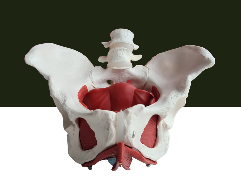
The pelvic floor is a group of muscles that supports the bladder, bowel, and sexual organs. These muscles also play an important role in erections, ejaculation, continence, and pelvic stability. When they become excessively tense or poorly coordinated, they can contribute to pain, reduced erectile quality, and discomfort during intimacy.
Patients often describe symptoms such as:
- Pelvic or perineal pain
- Pain during erections
- Pain during or after intercourse
- Difficulty achieving or maintaining a firm erection
- A persistent feeling of tightness in the pelvis
- Increased anxiety surrounding sexual activity
Many also say things like:
“Everything feels tight.”
“My erections don’t feel as full as they used to.”
“The curve seems more noticeable now.”
In these situations, the anatomical curve may not have changed at all. Instead, muscle tension, altered movement patterns, and increased nervous system sensitivity are amplifying symptoms.
This distinction is important because it changes how treatment is approached. A structural problem requires medical evaluation, while muscular dysfunction often responds well to conservative rehabilitation.
Bottom line: Pelvic floor dysfunction does not create a penile curve, but it can increase pain, worsen erectile function, and make an existing curve feel more symptomatic.
Why Does the Pelvic Floor Affect Sexual Function?
Many people think erections depend only on blood flow. In reality, healthy sexual function relies on the coordinated interaction of muscles, nerves, blood vessels, connective tissue, and the brain.
When the pelvic floor remains chronically tense, several things can happen:
- Muscles lose their ability to relax and contract efficiently.
- Blood flow may become less effective during arousal.
- Breathing mechanics become less efficient, increasing abdominal and pelvic tension.
- Chronic pain can heighten nervous system sensitivity, making otherwise mild symptoms feel more intense.
- Anxiety about sexual performance often reinforces muscle guarding, creating a cycle that is difficult to break.
This is one reason men with chronic pelvic pain, high stress levels, or prolonged sitting frequently experience erectile or sexual symptoms even when no structural penile abnormality is present.
Rather than viewing the penis in isolation, I evaluate how the entire lumbopelvic system functions together. Sexual health is influenced by movement, posture, breathing, hip mobility, abdominal control, and nervous system regulation—not just the anatomy of the penis itself.
Can Pelvic Floor Physical Therapy Straighten a Curved Penis?

This is one of the most common questions I receive.
The answer is no.
Pelvic floor physical therapy cannot straighten a penis with congenital curvature or Peyronie’s disease because those conditions involve structural anatomy that exercise or manual therapy cannot change.
Being clear about this expectation is important from the beginning. Physical therapy is not a treatment for scar tissue within the penis or developmental curvature.
What it can do is address the muscular and functional impairments that often accompany those conditions.
Treatment may help improve:
- Pelvic floor muscle overactivity
- Pelvic pain
- Painful erections
- Pain during intercourse
- Hip and pelvic mobility
- Breathing mechanics
- Core muscle coordination
- Movement patterns that increase pelvic stress
- Confidence during sexual activity
- Erectile function when pelvic floor dysfunction is contributing
For many men, these improvements have a meaningful impact on quality of life. Although the anatomical curve remains unchanged, erections become more comfortable, pelvic pain decreases, and sexual activity feels less restricted.
My goal is not to change your anatomy. My goal is to help you achieve the healthiest, most comfortable, and most functional version of it.
Key takeaway: Pelvic floor physical therapy improves function—not structure. While it cannot straighten the penis, it can often reduce pain, improve movement, and restore confidence when pelvic floor dysfunction is contributing to symptoms.
Why a Whole-Body Assessment Matters
One of the biggest mistakes in men’s pelvic health is assuming every symptom originates in the penis.
In practice, sexual function reflects the coordinated performance of the entire body.
During an evaluation, I assess far more than the pelvic floor alone. Depending on the patient’s presentation, this may include:
- Breathing mechanics
- Rib cage and diaphragm mobility
- Core muscle coordination
- Hip range of motion
- Lumbar spine movement
- Pelvic alignment
- Posture and movement patterns
- Exercise habits
- Occupational demands
- Stress and nervous system regulation
Restricted hip mobility, prolonged sitting, previous orthopedic injuries, abdominal muscle overactivity, and chronic stress can all increase tension throughout the pelvic floor.
For many patients, addressing these contributing factors leads to meaningful improvements in pain and sexual function, even though the structural curvature itself does not change.
This whole-body perspective also helps explain why two men with similar degrees of curvature can have very different experiences. One may have no symptoms at all, while another develops pelvic pain, erectile dysfunction, or difficulty with intercourse because additional muscular or biomechanical factors are involved.
Rather than treating the curve in isolation, rehabilitation focuses on improving the systems that influence comfort, movement, and sexual function as a whole.
When Is Pelvic Floor Physical Therapy Appropriate?
Pelvic floor physical therapy is most helpful when muscular dysfunction is contributing to symptoms alongside—or independent of—a penile curve.
You may benefit from an evaluation if you experience:
- Pelvic or groin pain
- Pain during erections
- Pain during or after intercourse
- Erectile dysfunction without a clear vascular cause
- Persistent pelvic floor tightness
- Lower abdominal or perineal discomfort
- Hip stiffness that affects pelvic movement
- Anxiety-related muscle guarding
Treatment begins with a comprehensive one-on-one assessment to identify the factors contributing to your symptoms. Rather than assuming the curvature is the sole cause, the evaluation examines how your muscles, joints, breathing, and nervous system interact to influence sexual function.
For some men, rehabilitation becomes an important part of recovery. For others, it complements medical management by improving the muscular and functional issues that remain after a diagnosis has been established.
Bottom line: Pelvic floor physical therapy is not designed to correct structural penile curvature. Its role is to identify and treat the muscular, movement-related, and nervous system factors that may be contributing to pain, erectile dysfunction, or limitations in sexual function.
When Should You See a Urologist?
Although many cases of penile curvature are harmless, certain symptoms warrant prompt evaluation by a urologist, who treats both sexual concerns and urinary problems. Early assessment is particularly important if the curve is new, worsening, or accompanied by pain or erectile dysfunction.
Seek medical evaluation if you notice:
- A curve that develops suddenly
- Progressive worsening of an existing curve
- Pain during erections
- A firm lump or plaque within the penis
- Significant penile shortening or narrowing
- Difficulty achieving or maintaining erections
- Blood in the urine or semen
- Trauma to the penis during sexual activity
- Severe pain or swelling after an injury
A urologist may perform a physical examination, or physical exam, and, when appropriate, recommend imaging such as penile ultrasound to evaluate for scar tissue or other structural abnormalities. Proper assessment may also involve examining the penis shaft and, when needed, the erect penis to document curvature; X-rays are standard for measuring the angle of curvature.
Depending on the diagnosis, treatment options may include observation, oral medications for symptom management, traction devices that gently stretch tissue over time, injectable therapies for selected cases of Peyronie’s disease, or surgical correction when severe curvature significantly interferes with sexual function.
Pelvic floor physical therapy can play an important complementary role, but it should never delay medical evaluation when warning signs are present.
Bottom line: If your curve is new, changing, painful, or affecting erections, see a urologist. Early evaluation helps determine the cause and guide appropriate treatment.
How Pelvic Floor Physical Therapy Fits Into Your Care
Many men assume they need to choose between seeing a urologist and seeing a pelvic floor physical therapist. In reality, the best outcomes often come from coordinated care.
Each provider addresses a different aspect of the problem.
A urologist evaluates the structural health of the penis, diagnoses conditions such as Peyronie’s disease, and discusses medical or surgical treatment when necessary, since treating Peyronie’s disease may involve both medical and surgical decision-making depending on the stage and severity.
A pelvic floor physical therapist evaluates how muscles, joints, breathing mechanics, posture, and movement patterns may be contributing to pain, erectile dysfunction, or limitations in sexual function.
These approaches are complementary rather than competing.
For patients with muscular dysfunction, rehabilitation can improve comfort, restore movement, reduce pelvic floor overactivity, and support recovery alongside medical treatment.
My Advice to Men Who Are Worried About a Curved Penis
One of the biggest sources of anxiety I see is the belief that a penis must be perfectly straight to function normally.
That simply isn’t true.
Mild penile curvature is common, and for many men it never causes pain, erectile dysfunction, or difficulty during intercourse.
Instead of focusing on appearance alone, pay attention to how your body is functioning.
Ask yourself:
- Has the curve changed recently?
- Is it painful?
- Are my erections different than they used to be?
- Is my sex life becoming uncomfortable or difficult?
- Am I avoiding intimacy because of pain or anxiety?
Those answers are far more meaningful than comparing your anatomy to photos online or trying to estimate the angle of the curve.
If your symptoms are stable, painless, and not affecting your quality of life, reassurance may be all that’s needed.
If they are changing or interfering with your daily life, seeking professional evaluation is the best next step.
Remember, the goal isn’t achieving perfect symmetry. It’s maintaining comfortable, confident, and satisfying sexual function.
Key takeaway: Focus on function, not perfection. Most mild curves are normal, but changing or symptomatic curves deserve professional evaluation.
Conclusion
If you’ve been wondering, “How much curve is too much?” the answer isn’t determined by a specific number of degrees.
A mild, stable curve is a common anatomical variation and often requires no treatment. What matters is whether the curvature changes over time, causes pain, affects erections, or interferes with sexual activity.
Congenital penile curvature and Peyronie’s disease are different conditions with different causes and treatment approaches. Distinguishing between them is an important first step toward receiving appropriate care, especially since the exact cause of Peyronie’s disease is not always known and some men develop Peyronie’s disease after an injury or repeated microtrauma.
Pelvic floor physical therapy cannot straighten a structurally curved penis. However, it can be highly effective for addressing the pelvic pain, muscle tension, movement dysfunction, and erectile difficulties that may accompany a penile curve or occur independently.
If you’re experiencing new or worsening curvature, painful erections, or difficulty with sexual function, don’t rely on internet forums or self-diagnosis. A comprehensive evaluation can help determine whether your symptoms are related to normal anatomy, Peyronie’s disease, pelvic floor dysfunction, or a combination of factors.
With the right diagnosis and a coordinated treatment plan, most men can improve their comfort, confidence, and quality of life, although research has not identified a reliable way to prevent Peyronie’s disease.
Frequently Asked Questions
Is a slightly curved penis normal?
Yes. A slightly curved penis with a slight bend is common and usually represents a normal anatomical variation. Treatment is generally unnecessary unless the curve is painful, worsening, or interferes with sexual function.
What degree of curvature is considered too much?
There is no universal cutoff. Although research often references 30 degrees when discussing treatment, the decision to seek evaluation should be based on symptoms rather than angle alone. Pain, progression, erectile dysfunction, and difficulty with intercourse are more clinically important than the degree of curvature.
Can pelvic floor dysfunction cause a curved penis?
No. Pelvic floor dysfunction does not cause structural penile curvature. However, it can contribute to pelvic pain, painful erections, reduced erectile quality, and muscle tension that make an existing curve feel more symptomatic.
Can pelvic floor physical therapy straighten my penis?
No. Physical therapy cannot correct congenital curvature or the scar tissue associated with Peyronie’s disease. Its role is to improve muscle function, reduce pain, optimize movement, and address pelvic floor dysfunction that may be contributing to your symptoms.
How do I know if I might have Peyronie’s disease?
Peyronie’s disease is more likely if the curve develops later in life, worsens over time, causes painful erections, is associated with a firm plaque beneath the skin, or leads to penile shortening or erectile dysfunction. Key risk factors include age, family history, and connective tissue conditions. These symptoms should be evaluated by a urologist.
When should I seek professional help?
You should seek evaluation if you experience a new or worsening curve, painful erections or injury during sexual intercourse, erectile dysfunction, difficulty with intercourse, a palpable lump, or significant anxiety about changes in your penile shape or function.
Ready to Get Answers?

If you’re concerned about penile curvature, pelvic pain, erectile dysfunction, or changes in sexual function, a comprehensive evaluation can help identify the factors contributing to your symptoms.
At Pelvis NYC, we take a whole-body approach to men’s pelvic health. Evaluating not only the pelvic floor but also the hips, core, breathing mechanics, posture, and movement patterns that influence sexual function. When appropriate, we coordinate care with trusted urologists. This ensures you receive the most effective treatment for both the structural and functional aspects of your condition.
Whether your symptoms are related to a normal anatomical variation, pelvic floor dysfunction, Peyronie’s disease, or a combination of factors, our goal is to help you understand what’s happening, reduce pain, improve function, and return to comfortable, confident intimacy.
👉 Book Now: https://calendly.com/drpelvis/appointment